Back to top: A16 Burials
I. Late Khabur Burials
Back to top: A16 Burials
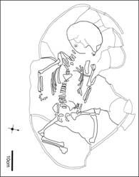
I.1 Introduction to A16.31 (a9)
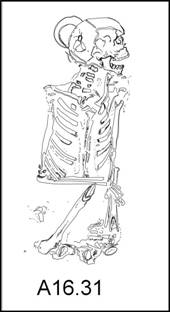
A16.31 are the remains of an adult female between 25-35 years of age, dating to the late Khabur period. She was buried within an area identified as a cemetery and recovered in primary context, inside a subterranean vaulted mud-brick tomb built alongside tomb a6, reusing the same wall. Small traces of gypsum were found inside the tomb’s walls, indicating that the tomb was plastered inside. A bronze bucket shaped vessel labeled as i29, was found at the same level where the walls of the tomb were first identified, approximately 80 cm above the floor of the tomb. Emplacement near the top of the tomb suggests that the bucket either hung from the tomb’s ceiling or was placed above the tomb. The burial chamber was accessed through the eastern face via a small corridor with the doorway found lined or blocked by three stones.
The body was recovered resting on her left side in a flexed position oriented north (head) to south (pelvis) found in good preservation state however her lower legs and feet were not recovered. The absence of the lower legs from the distal end of the femora shafts suggests that the ligaments were still in place to keep the foot and leg articulated and must have been removed before the body was fully skeletonized. No animal gnawing marks were found on the femora to indicate animal activity in carrying off the lower legs however the left femur was found disarticulated, located near the western tomb wall. Another possibility besides animal disturbance is post-mortem removal of the lower legs for secondary burial or ritual of which much evidence exists at Mozan for post-mortem body manipulation.
Osteological analysis found that A16.31 was a petite female between 4’8-5’1, using the ulna for stature estimation since the femora were incomplete. She suffered from an inflammatory condition of the teeth and jaw as evident by a large periapical abscess on her upper left first molar caused by a cavity that spread to the root and gum. Pressure from the inflammation would have caused intense pain and discomfort as the bony gum developed a fistula to drain the excess production of pus and may have resulted in her death if the infection spread. Her bones have rugged muscle attachment points, indicating she regularly participated in strenuous activity, particularly movements associated with the arms, common among the inhabitants of Urkesh at this time. Other observations of note is evidence for bilateral woven bone in the acetabulum, the hip socket for the leg, and small clusters of pin-like holes in the inner table of the cranium. No other joints are affected. These conditions are most likely normal variations and without a complete vertebral column and lower leg, differential diagnosis is not possible.
Back to top: A16 Burials
I.2 Preservation
In good condition; 61% post cranium, 95% cranium, and 100% of ribs complete. The lower legs are missing including the tibiae, fibulae, and feet. Teeth are in excellent condition although six are missing post-mortem.
Back to top: A16 Burials
I.3 Biological Age
Biological age was estimated through various aging techniques. Dental wear visible on the mandibular molars indicates moderate wear with the cusps on the buccal side showing heavier wear on the first molar. Using Miles 1962 attrition scheme, age estimation through dental wear is within range 28-36 years. Age estimation through deterioration of the pubic symphysis found that the left symphysis displayed visible billowing present throughout most of the surface, becoming less defined in the upper extremity eventually replaced by ossification nodules. There is little distinction between the dorsal and ventral surfaces, no dorsal plateau present, nor lipping. Given these characteristics, age estimated as stage 2: mean age 25+/-4.9 (Brooks and Suchey 1990). The right pubic symphysis shows a slightly older individual with a distinction between the ventral and dorsal surfaces as the dorsal develops a plateau and loss of billowing. The top of the symphyseal face is broken but ossification nodules are present. The right symphyseal phase is estimated as stage 3: mean age 30.7 +/-8.1. Combined estimations for the pubic symphysis are within the range of 23-35 years of age with an average of 25-35 years when the Miles attrition scheme is factored in.
Back to top: A16 Burials
I.4 Biological Sex
Biological Sex was determined using sexually diagnostic characteristics of the pelvis and skull. While the skull was found to contain several typical male traits such as a robust jaw and large mastoid process, the pelvis was found to be predominately female in range with the presence of a ventral arc, sub-pubic concavity, preauricular surface, U-shaped greater sciatic notch, and a broad pelvis placing A16.31 well within the female range.
The following two charts summarize the characteristics noted.
|
|
||
|
Overall shape (anterior view) pelvis |
low and broad |
Female |
|
Greater sciatic notch |
U-shaped, obtuse |
Female |
|
Auricular surface |
narrow on elevated plane |
Female |
|
Preauricular sulcus |
present |
Female |
|
Acetabulum |
small, faces anterolaterally |
Female |
|
Pubic symphysis height |
Tall |
Male |
|
Pubic rami |
long |
Female |
|
Sub-pubic angle |
broad and U-shaped |
Female |
|
Pubic tubercle |
sharp, further from symph. |
Female |
|
Inferior pubic ramus |
gracile, tapers superiorly |
Female |
|
Ventral arc |
present |
Female |
|
Sub-pubic concavity |
present |
Female |
|
obturator foramen |
large, tall, ovoid |
Male |
|
Width of sacral ala |
Broader than body of S1 |
Female |
|
Anterior sacral curvature |
Extends from S3-S5 |
Female |
|
|
||
|
Overall shape of cranium |
gracile |
Female |
|
Glabellar profile |
Smooth |
Female |
|
Frontal slope |
vertical |
Female |
|
Frontal and parietal tuberosities |
reduced |
Male |
|
Zygomatic process of frontal |
narrow |
Female |
|
Supraorbital ridges |
slight |
Female |
|
Nasal bone |
small |
Female |
|
zygomatic bone |
short |
Female |
|
Suprameatal crests |
short |
Female |
|
Mastoid process |
largel |
Male |
|
Nuchal area |
ridged |
Male |
|
External occipital protuberance |
small |
Female |
|
Occipital and mandibular condyles |
small |
Female |
|
Canine eminence |
indistinct |
Female |
|
Palate |
small |
Female |
|
Mandibular ramus (ant-post) |
In-between |
Male? |
|
Depth from incisor to mentum |
short |
Female |
|
Mental protuberance |
large |
Male |
|
Lower margin of mandibular corpus |
thin |
Female |
|
Angle of mandible |
obtuse |
Female |
|
Lower first molar |
four cusped |
Female |
Back to top: A16 Burials
I.5 Stature
Estimated at145-154 cm (4’8-5’1) using Trotter and Gleser 1952; formulae for “negro females” using a complete ulna measuring 242 mm.
Back to top: A16 Burials
1.6 Pathology and Health
Tooth abscess, periodontal disease, enamel hypoplasia, and carious lesions noted. Teeth are in excellent condition and display slight calculus build-up with medium periodontal disease. Carious lesions were visible on LM1 affecting the buccal root surface, LM2 at the enamel root junction on the distal end, and RM1 lingual side where the cavity developed into a large abscess on the buccal surface near the nasal cavity. The lesion shows smooth surfaces with periodontal disease noted on the sequential molars with the gum recessed and inflamed. The cervical vertebrae exhibit slight lipping, increased porosity, and Schmorl’s nodes. Little of the thoracic and lumbar vertebrae were recovered due to their poor condition however the fragments available do suggest increased porosity and osteophyte development. The right acetabulum contains woven bone formation with holes on the acetabular fossa that does not extend into the lunate surface. The region with the most activity is at the center of the fossa, away from the lunate surface. Similar woven bone formation is also visible on the left acetabulum however the porosity is immediately next to the lunate surface on the lateral end of the fossa. On the cranium, clusters of small pin-like holes varying in size were noted on the cruciform eminence of the inner occipital bone. Only the inner table is affected with no changes visible on the outer surface of the cranium.
Stress Markers (MSM) visible on both
deltoid tuberosities (slight) with the right humerus showing a deep
groove for the long head of Biceps brachii. On the ulna the supinator
crest shows ruggedness with more pronounced points on the right side
as well as MSM points on both linea aspera of the femur with the left
more rugged.
Photos: 19 views
VDS70l TA A16.31 Pubic bones, ventral side displaying a prominent pubic tubercle.
VDR824 TA A16.31 Pubic bones, dorsal view showing pubic concavity, indicating this is a female.
VDR824 TA A16.31 Left pubic symphysis.
VDR824 TA A16.31 close-up, abscess on maxilla, left side first molar.
VDR824 TA A16.31 dental cavity on root surface, left first mandibular molar.
VDR824 TA A16.31 left pubic symphyseal face.
VDS703 TA A16.31 right pubic symphyseal face.
VDS701lr Left pubic symphyseal face at an angle to show billowing surface.
VDR824 TA A16.31 dental cavity affecting gum, left maxilla.
VDR824 TA A16.31 dental abscess on the right maxilla.
VDR824 TA A16.31 mandibular molars, right side.
VDO830 lr A16.31 (1) Mandible, view from right.
VDR824 TA A16.31 close-up of occipital, inner table view.
VDR824 TA A16.31 view of vertebral body with schmorl’s nodes.
VDR824 TA A16.31 left acetabulum.
VDS701lr cranium inner view after reconstruction.
VDR824 TA A16.31 occipital, inner table view cruciform eminence with possible pathology.
VDS701lr cranium outer after reconstruction.
VDR824 TA A16.31 cranium.
Back to top: A16 Burials
II. Early Khabur Burials
Back to top: A16 Burials
II.1 Introduction to A16.123 (a20)
These are the skeletal remains of a small child between 5-12 months of age, with a non-specific infection affecting the outer bone layer of the skeleton. The child was found wearing bronze circular hooped earrings, lying supine over two large sherds from a ceramic vessel. The sherds were reconstructed after excavation, forming an incomplete large ceramic jar with a convex base, incised with three lines below the neck. Another burial was found within close proximity, A16.156, located twenty-five centimeters below A16.123 and slightly north. This tomb contained a mud brick wall to demark the burial which was exposed at the same elevation as infant burial A16.123. Given the close proximity of these two burials it is possible that they are associated with each other. A16.156 was estimated as a female between 22-30 years, perhaps a mother with her child that died at a later date.
Back to top: A16 Burials
II.2 Biological Age
Infant, estimated within the range 5-12
months of age via multiple aging techniques. The diaphyseal length of
the long bones are as follows: Humerus 66mm, Ulna 85mm, radius 74mm,
tibia 95mm, femur 113 mm. Aging via the diaphyseal length utilizing
Hoppa’s 1992 method, suggests an age of 12 months. Bone
ossification suggests a slightly younger age at 9-12 months based on
the presence of a fused mandible (>6-9 months) while the vertebral
bodies remain un-fused (<12 months) (Buikstra and Ubelaker 1994).
Deciduous teeth formation gives an estimate within 5-10 months based
on: canine at stage Crc= 5-8 months, molar 1 at stage r1/4= 5-10
months, and permanent molar 1 at Coc= 6 months (Moorees, Fanning and
Hunt 1963; Smith 1991).
|
Ossification/Fusion stage {#} |
Age range {#} |
|
mandible fused |
>6-9 mo |
|
vertebral bodies unfused |
<12 mo |
|
Bone {#} |
lg/mm {#} |
age {#} |
|
Humerus |
86 |
12 mo |
|
Ulna |
85 |
12 mo |
|
Radius |
74 |
12 mo |
|
Tibia |
95 |
12 mo |
|
Femur |
113 |
12 mo |
|
Tooth {#} |
Stage {#} |
Age {#} |
|
M1 |
Coc |
6 mo |
|
Dc |
Crc |
5-8 mo |
|
dm1 |
r¼ |
5-10 mo |
Back to top: A16 Burials
II.3 Preservation
The bones were found in good condition with 43% post cranium, 85% skull, and <75% of the ribs complete.
Back to top: A16 Burials
II.4 Pathology and Health
The left ulna and fibula show signs of periostitis, an inflammation of the periosteum either through trauma or infection, with evidence of woven bone formation on the shaft that takes on a porous appearance. The inflammation is nonspecific resulting from either periostitis as the primary disease or as a secondary reaction of another disease. No other skeletal lesions where noted. In archaeological skeletal material, periosteal inflammation of the long bones forms the most common pathological condition recorded (Ortner 2003).
Measurements of the Ulna: 85 mm; radius 75 mm, tibia 95 mm, and femur 113 mm.
No photographs of the skeleton from lab.
Back to top: A16 Burials